
|
Erythroplakia
Erythroplakia. Note the red patch on the right buccal mucosa with white areas posteriorly.
|
|
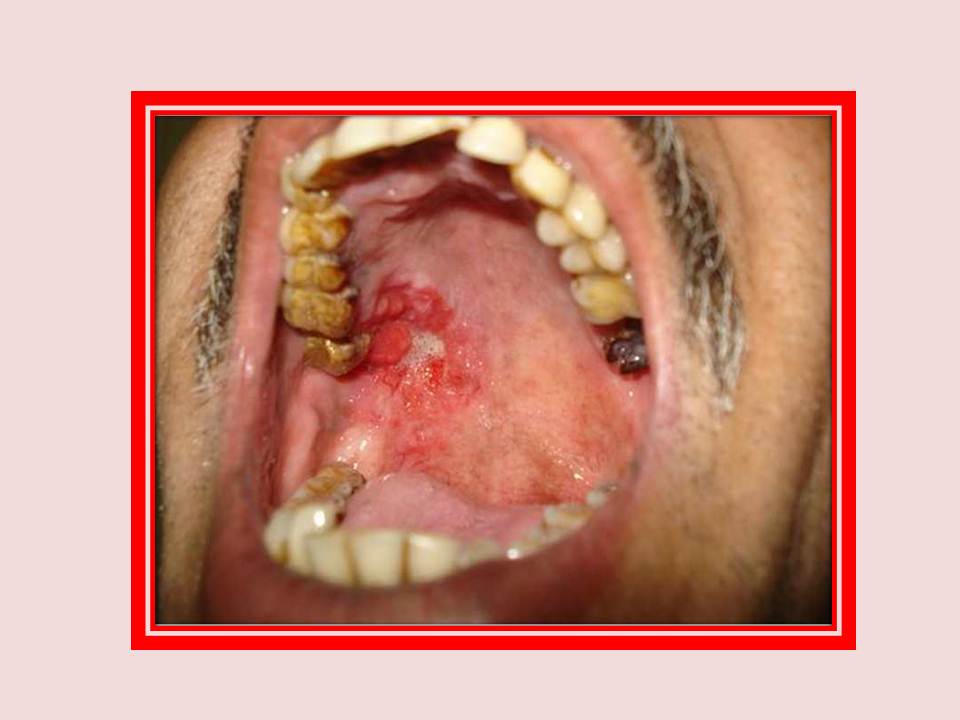
Erythroplakia
Erythroplakia. Note the red velvety lesion involving the posterior aspect of the right side of the hard palate. Note the raised irregular area along the anterolateral aspect of the lesion which is clinically suspicious of malignant transformation.
|
|
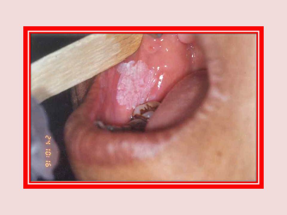
Leukoplakia
Homogeneous leukoplakia on the right buccal mucosa.
|

|
Leukoplakia
Nodular leukoplakia on the right buccal mucosa. A well-circumscribed lesion of 3x1 cm size on the right buccal mucosa in a 63-year-old habitual betel quid chewing female. Note the pin head sized nodules scattered on an erythematous base.
|

|
Leukoplakia and Candidiasis
Figure A: Superadded candidiasis in a patient with homogeneous leukoplakia.
Figure B: The same patient after three weeks of antifungal treatment.
|

|
Leukoplakia
Proliferative verrucous leukoplakia (PVL). Note the extensive, thick, white plaques.
|
|
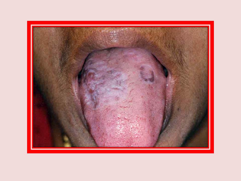
Lichen planus
Lichen planus. Note the white 4x3.5 cm patch on the right side of the dorsum tongue intermingled with areas of pigmentation. Another annular form of lichen planus can be seen on the left side of the dorsum tongue.
|
|
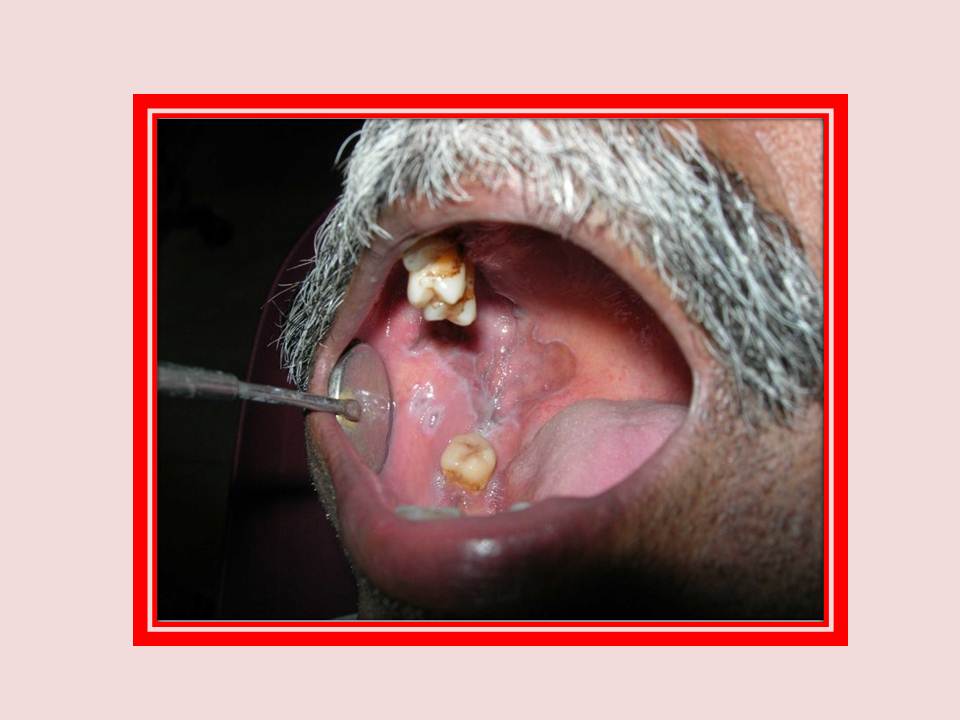
Lichen planus
Annular lichen planus. Note the ring-like pattern on the right buccal mucosa, retromolar trigone and hard palate.
|
|
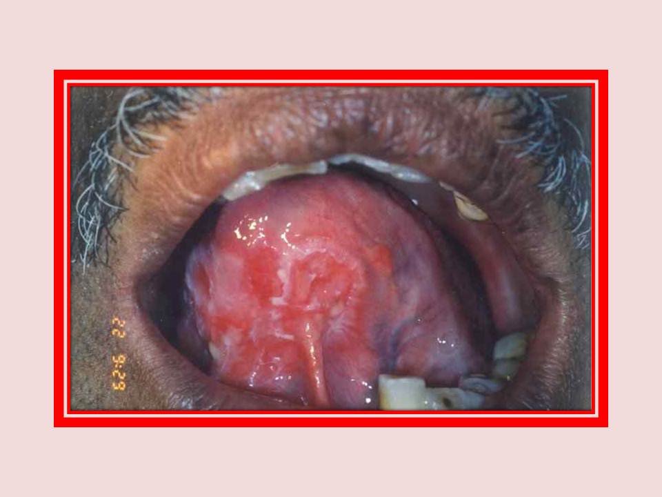
Lichen planus
Erosive lichen planus on the ventral surface of the tongue, just above the lingual frenum, surrounded by white striae.
|

|
Oral submucous fibrosis
Oral submucous fibrosis. Note the blanching on the lower labial mucosa.
|

|
Oral submucous fibrosis
Oral submucous fibrosis with erythroplakia of the tongue.
|
|
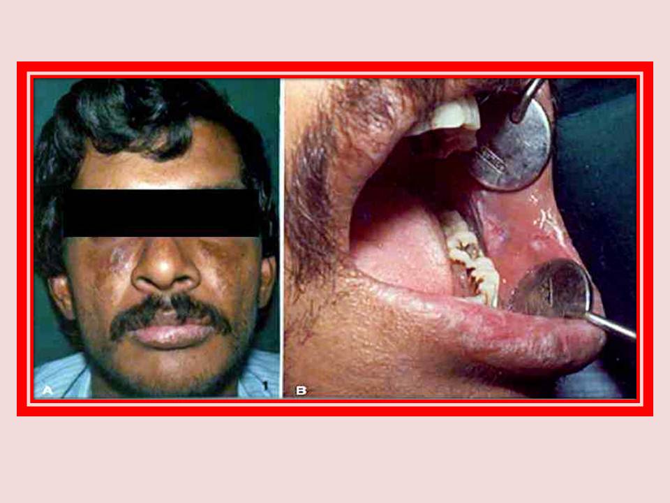
Lupus erythematosus
Figure A: Extra oral photograph of a patient with discoid lupus erythematosus. Note the butterfly-shaped rash on the malar area.
Figure B: Intraoral photograph of the same patient showing erosive lesions surrounded by radiating white striae on the left buccal mucosa.
|
|
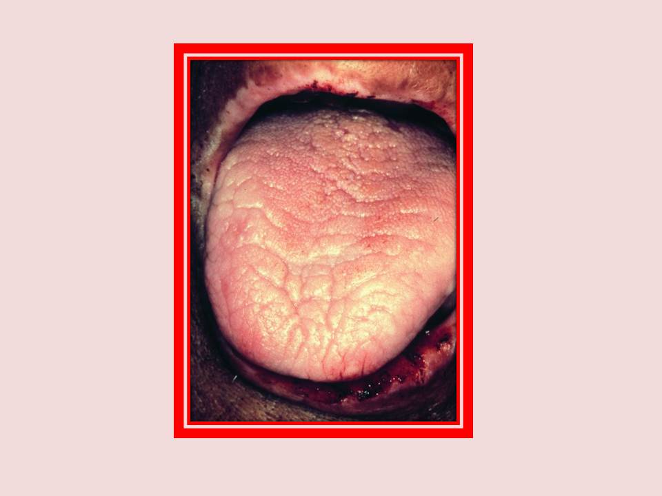
Sideropenic dysphagia
Sideropenic dysphagia. Iron deficiency anaemia with depapillated tongue, depigmentation of the upper lip and epithelial erosion of the lower lip.
|
|
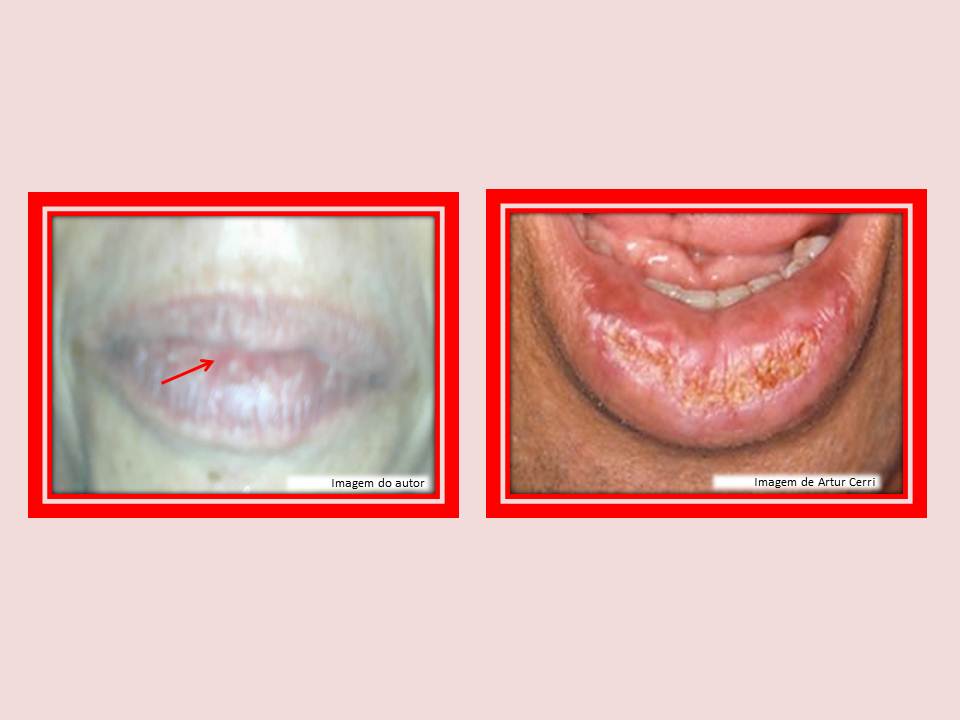
Actinic Cheiloses
Actinic Cheiloses. Note the actinic keratosis affecting the lip vermilion. Usually happens as a result of too much sun exposure.
|
|
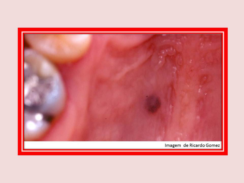
Nevus
Observe the presence of pigmented lesion of recent growth, irregular margins, and variable color. The presence of ulceration and satellite lesions raise the possibility of melanoma.
|
|
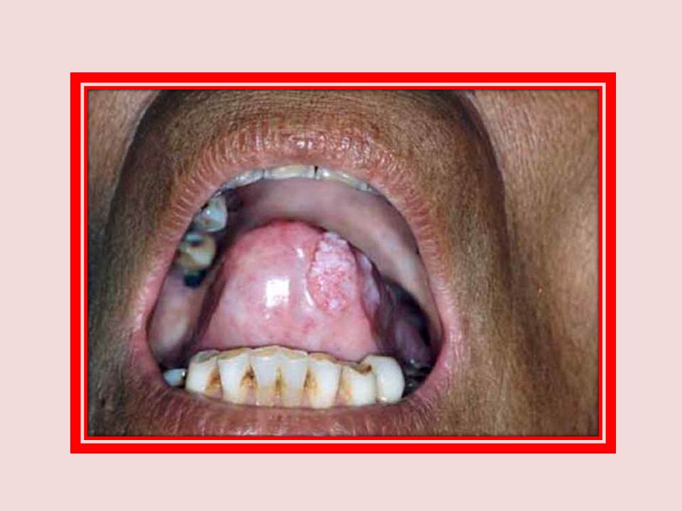
Oral submucous fibrosis
Oral submucous fibrosis with malignant growth on the left side of the anterior part of the oral tongue.
|
|
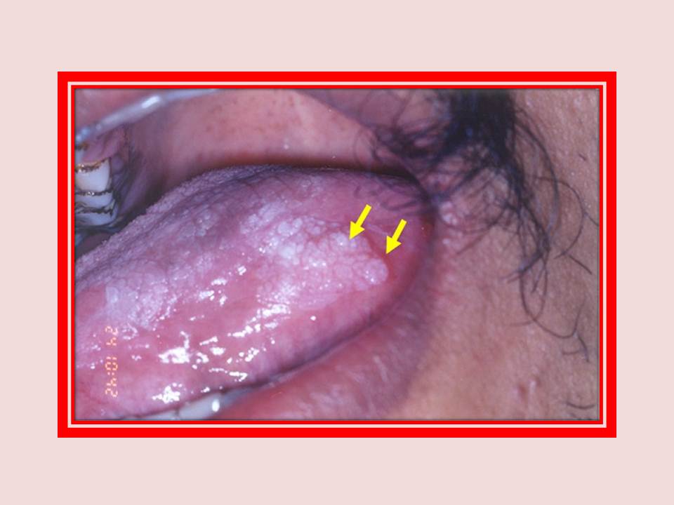
Leukoplakia
Homogeneous leukoplakia on the dorsum and left lateral margin of the tongue, showing malignant transformation. Note the raised, erythematous posterior margin of the white plaque (arrows). Well-differentiated squamous cell carcinoma was established on histopathological examination.
|
|
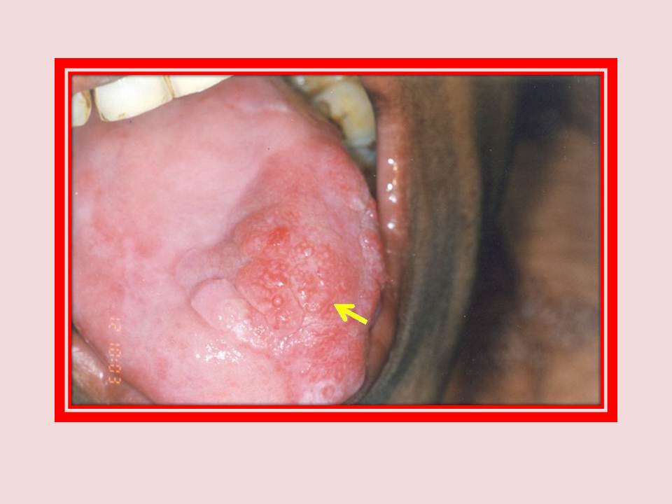
Oral submucous fibrosis
Oral submucous fibrosis with a nodular, proliferative growth on the anterior aspect of the dorsum of the tongue. Well differentiated squamous cell carcinoma was established on histopathological examination.
|